
Percutaneous Fixation Using Buck’s Technique:
A Minimally Invasive Solution for L5 Pars Fractures
Clinical insights from a case series of 13 patients treated with the FacetFuse system by KIC Ventures
- Enrique Azmitia, PhD a, Mario Cahueque, PhD b,*
a Department of Neurosurgery, Hospital Herrera Llerandi, Guatemala, Guatemala
b Department of Orthopedics and Traumatology, Hospital Centro Médico, Guatemala, Guatemala
- Anshul Jain
Founder’s Office, KIC Ventures

Introduction
Pars interarticularis fractures at the L5 level are a relatively uncommon but important cause of chronic lower back pain, particularly in young, active individuals. These fractures often result from repetitive hyperextension or rotational stress, making athletes in sports like football, volleyball, gymnastics, ballet, and CrossFit especially susceptible.
While conservative treatment is typically the first line of management, a subset of patients fails to improve after extended periods of rest, physical therapy, and bracing. For these cases, surgical intervention is indicated. Yet, there remains limited clinical data on the outcomes of percutaneous repair using Buck’s technique. A recent retrospective case series published in Advanced Spine Journal (2025) addresses this gap, documenting the experience of 13 patients who underwent this technique using FacetFuse cannulated screws (4.0 mm) from KIC Ventures.
Study Summary
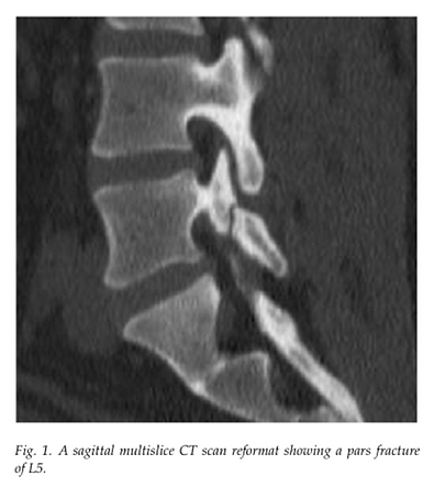
The study focused on 13 patients, aged 19 to 42 years (average age 28), all of whom had symptomatic L5 pars fractures confirmed on imaging and had failed at least six months of conservative care. Importantly, none had associated spondylolisthesis, making them ideal candidates for direct fracture repair.
All patients were treated using Buck’s percutaneous fixation technique under fluoroscopic guidance. The procedure involved precise bilateral guidewire placement across the fracture line, followed by insertion of 4.0 mm cannulated screws through small incisions. The use of fluoroscopy ensured accurate screw trajectory with minimal tissue disruption.
Postoperatively, patients were mobilized on the same day, with high-impact activities restricted for three months.
Key Results
Patients reported significant improvements in pain, measured via the Visual Analog Scale (VAS). The average preoperative VAS score was 6.5. At six weeks post-op, pain levels dropped by nearly 40 percent. By three months, the average pain reduction reached 69 percent, and by one year, patients experienced over 80 percent improvement. These outcomes were maintained at two-year follow-up.
CT imaging confirmed bony union in all cases by six months. There were no reported intraoperative complications, infections, hardware failures, or nonunions. Nearly all patients returned to their previous level of activity within a year, including resumption of competitive sports.
Why This Matters for Spine and Ortho Physicians
This study highlights several clinical takeaways:
-
Minimally invasive repair is a viable alternative to fusion for select patients with isolated pars defects.
-
The technique preserves spinal motion and avoids the morbidity associated with segmental fusion.
-
With modern instrumentation like FacetFuse, percutaneous Buck’s technique can be executed with precision, reproducibility, and high safety margins.
-
Young athletes or active patients can benefit from shorter recovery times and a quicker return to function.
For surgeons seeking motion-preserving solutions for symptomatic pars fractures, this case series demonstrates that direct percutaneous fixation can be both effective and durable when conservative options fail.
Elevating Standards Through Research
We extend our sincere thanks to the authors of this paper, Enrique Azmitia, PhD and Mario Cahueque, PhD, for their important contribution to the field. By sharing real-world data on percutaneous Buck’s technique using the FacetFuse system, they are helping advance motion-preserving care for active patients with L5 pars fractures. Their work not only fills a critical gap in the literature but also empowers surgeons with evidence to support better clinical decisions.
Conclusion
Buck’s technique, performed percutaneously with current-generation implants, offers a safe, effective, and motion-preserving option for patients with chronic L5 pars fractures. The FacetFuse system enhances procedural accuracy and stability, making this approach increasingly accessible to spine and orthopedic surgeons.
To learn more about this technique or to explore peer training opportunities, contact KIC Ventures for further information.